
Al for Thyroid
Nodule Detection


1 in 4-5
1 in 4-5 people are affected by thyroid nodules
Source: American Thyroid Association, Johns Hopkins Medicine, National Center for Biotechnology Information
30%
Up to 30% of thyroid nodules are indeterminate cases by conventional ultrasound interpretation.
Source: American Thyroid Association, Mayo Clinic
3x
Thyroid cancer cases have tripled over the last decade.
Source: American Cancer Society, National Cancer Institute
5th
Thyroid cancer ranks among the top 5 most common cancers in women in the USA (5th), Taiwan (5th), China (4th), and South Korea (1st).
Source: American Cancer Society and National Cancer Institute

Thyroid Workflow Made Simple
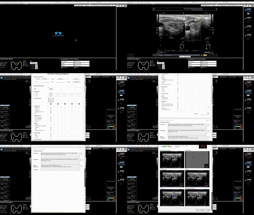
Automatic Nodule Detection & Contouring
Automatically identifies and outlines nodules as soon as the image is loaded. No manual tracing needed.
1
2
Instant TI-RADS Analysis
Extracts key sonographic features and provides a TI-RADS score within seconds. Fast, consistent, and objective.
3
Automated Report Generation
A diagnostic report is automatically created, ready for review and export

Advanced AI Capabilities

Color-Coded Nodule Features
Visualizes key characteristics such as echogenicity, shape, margins, and calcifications using patented visualization technology.
Support 8 TI-RADS Guidelines
Supports 8 major TI-RADS systems. Select your preferred guideline for assessment. The report can include results based on the selected system across others.
Quantified Nodule Sonographic Characteristics
Quantifies malignancy level of key sonographic features using patented algorithms.
Cross Platform Flexibility
Supports B-mode ultrasound images analysis on Android and Windows platforms.
Validated Clinical Accuracy
AI raises accuracy for readers of all experience levels
Parametric average ROC curve

0
0.2
0.4
0.6
0.8
1
Sensitivity
0
0.2
0.4
0.6
0.8
1
1 - Specificity
Overall AUC: +8.8%(Junior 0.78 → 0.85)
Reader-to-reader variability: -14%
Sensitivity: +30%
Specificity: +55%
Average AUC with and without AmCAD-UT of junior and senior group.
without CAD (junior)
with CAD (junior)
without CAD (senior)
with CAD (senior)

How-to Videos
AmCAD-UT for Handheld Ultrasound
Real-time AI Detection
Use your existing handheld ultrasound device with our real-time AI assistant

AmCAD-UT DICOM Analysis on PC
Automated AI Analysis of Any Thyroid Ultrasound Image
Analyze DICOM files from any ultrasound machine. Fast, precise, and PACS compatible.

